Dental X-Rays in Bali: Periapical, Panoramic, and CBCT Explained
Dental X-rays show structures that cannot be fully assessed during a visual examination, including roots, bone, and some hidden decay. The imaging…
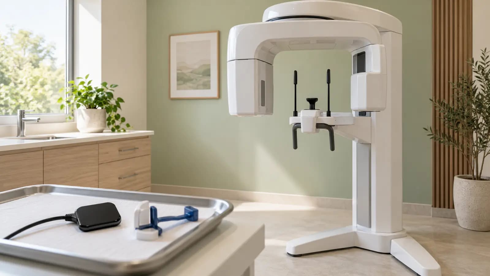
Technique should answer a clinical question. A three-dimensional CBCT scan is not automatically better for every patient.
If you need dental X-rays in Bali, discuss the purpose first and bring previous images where available. Your dentist can assess whether those records are sufficient or additional imaging is justified.
How the main techniques differ
Periapical images
A periapical image shows one or a few teeth from crown to root, including nearby tissues. It can help assess a specific area or support root canal diagnosis and treatment.
The sensor is usually placed inside the mouth. Tell the team about a strong gag reflex or difficulty opening so positioning can be adapted where possible.
Panoramic images
A panoramic dental X-ray in Bali gives a broad two-dimensional view of teeth and jaws. The machine moves around the head while you remain in the required position.
A broad view can help some planning questions, but it does not provide the same detail for every task as an intraoral image. It may not replace periapical or bitewing imaging when a specific area needs closer assessment.
CBCT scans
Dental CBCT in Bali provides three-dimensional information for selected indications, such as implant planning or anatomical relationships that are unclear on two-dimensional images.
The scan needs a clinical reason. The field of view and settings should suit the question while limiting unnecessary exposure. It is not a routine test for every toothache.
Other techniques include bitewings for areas between teeth and cephalometric images for selected orthodontic needs. Dental imaging is not limited to only two or three options.
When is imaging needed?
Symptoms, examination findings, disease risk, previous records, and the proposed treatment influence the decision. A tooth can look intact externally while having a hidden problem, but this does not mean every patient needs images at every visit.
There is no universal interval suitable for everyone. You can ask what the dentist wants to investigate and how the result would change the treatment plan.
Safety and individual circumstances
X-rays use ionising radiation. The aim is to obtain necessary diagnostic information using an appropriate technique, area, and exposure.
Tell the team if you are pregnant or might be pregnant. Necessary imaging is not automatically prohibited, but the information is relevant to planning. Children’s examinations also need settings and indications suited to their size and needs.
Requesting extra scans simply to feel more thoroughly checked can create unnecessary exposure. Equally, discuss concerns before declining imaging that is important to diagnosis.
Preparing for the appointment
- Bring existing images and their dates if available.
- Explain symptoms, previous treatment, health conditions, and relevant pregnancy information.
- Follow instructions about removing metal items or accessories from the imaging area.
- Keep still in the required position to reduce blurred images.
- Ask how to obtain the results and who will explain them.
For international patients, copies of images and treatment notes can help your regular dentist understand what was done in Bali. The images still require clinical interpretation; a dark area alone is not a self-diagnosis.
Fees and access
Costs depend on the imaging technique, area, and interpretation requirements. Confirm the fee and location after the appropriate examination is recommended. A clinic may provide available imaging or refer to a suitable radiology facility.
Use imaging to support a clear decision
For dental care and a discussion with a dentist Bali about imaging, contact Floss Dental in Kerobokan. An initial assessment can identify the relevant investigation and the next step based on clinical findings.
FAQ Seputar Dental X-Rays
Why are dental X-rays needed?
Some root, bone, and hidden tooth problems cannot be assessed visually. The need is determined individually.
What are the two main groups of dental X-rays?
They are commonly described as intraoral and extraoral, with several techniques within those groups.
How much do dental X-rays cost?
Fees vary by technique and facility. Ask for an estimate for the recommended image rather than a generic scan package.